Taken from the book The Machinery of Life by David S. Goodsell
As we all know, the mitochondria is the powerhouse of the cell.
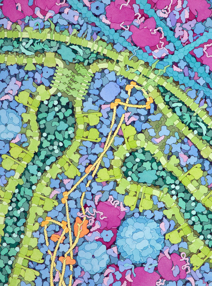
Unlike simpler bacterial cells, our cells are filled with compartments that perform different duties. ATP synthesis is the primary task performed by the mitochondria, shown here in cross-section. The enzymes of the citric acid cycle (purple & blue) are found in the innermost space and the folded inner membrane provides the necessary separation of spaces needed to create the electrochemical gradient that powers ATP synthase (green mushroom shape). This membrane is one of the most protein-concentrated membranes found in our cells: by some estimates, three quarters of the membrane is composed of protein, with just enough lipid to hold it together. Compare this illustration with the illustration of Escherichia coli in this previous post, and notice that the mitochondrion includes very similar machinery for protein synthesis, complete with DNA, RNA, and ribosomes. It does not make all of its own proteins, however, and some must be transported from the cytoplasm by specialized transport proteins (light green crown shape) in the inner and outer membranes (1,000,000 X)
More on the advanced compartmentalization of human cells and the odd origin of mitochondria:
The human body is composed of trillions of cells. Just like a bacterial cell, each one of our cells (with a few exceptions) has DNA, polymerases, and ribosomes for building proteins based on genomic information. Each cell is filled with a collection of enzymes for making and breaking the many molecules needed for growth and energy production. Each cell is surrounded by a sturdy cell membrane filled with channels, pumps, and sensors. Each of our cells, however, is much larger and far more complex than a bacterial cell. Where bacteria are built to be fast and efficient, our cells are made to perform complex and specific tasks, and they are made to last and perform these tasks for years.
By looking at the similarities and differences between bacterial cells and animal cells, scientists have discovered that a breakthrough occurred in the evolution of life about 1.5 billion years ago. Simple, bacteria-like cells had probably been around for at least 2 billion years, and most of the basic molecular machinery of life had been perfected. Then, a new design of the cell body appeared: larger cells filled with many small, separate compartments, each surrounded by its own watertight membrane. The proliferation of these new cells signaled a major break in the evolution of cells, and in the years since then, two distinct family lines have evolved. The simple cells, with their molecular machinery jumbled together in one compartment, are the distant ancestors of the bacteria and archaebacteria. The new compartmented cells gave rise to all other forms of life, including protozoa, fungi, plants, and animals.
Surprisingly, when we look under the microscope, some of these compartments look similar to whole bacterial cells. The mitochondria in each of our cells have a similar shape, size, and composition as an Escherichia coli cell. For instance, mitochondria are surrounded by two membranes. The outer membrane is studded with proteins reminiscent of bacterial porins. The inner membrane, pleated and folded in mitochondria, is filled with proteins of transport and energy production similar to the bacterial proteins. Even more surprising, when you look inside a mitochondrion, it has its own DNA, transfer RNA, and ribosomes, all busily constructing mitochondrial proteins. These remarkable similarities have prompted a theory of the origin of mitochondria (and chloroplasts) that is now widely accepted. It is thought that a bacterial cell took to living inside another cell in the distant past, perhaps entering as a parasite or perhaps surviving the process of being eaten. The bacterium continued to live in this comfortable, sheltered environment, living and reproducing inside the cell. Gradually, both the engulfed cell and the cellular host became increasingly reliant on each other, dividing up the tasks of living. Today, our mitochondria continue to divide and reproduce inside our cells, but they rely on the rest of the cell for many of their essential proteins and nutrients.
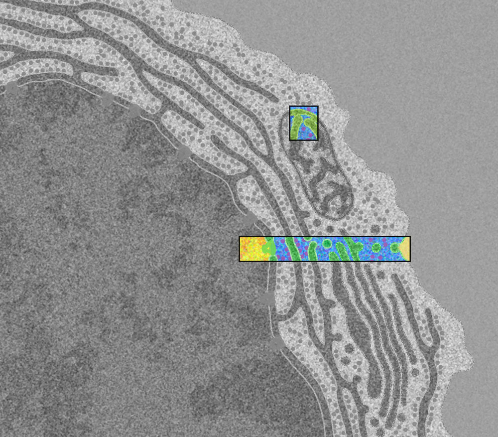
Here is a zoomed-out view (10,000 X) of a blood plasma cell under a microscope; the topmost box shows this post's headline illustration in context. The sheer scale of each ordinary human cell is incredible! And we have trillions of them!


